Chromosome Banding Patterns

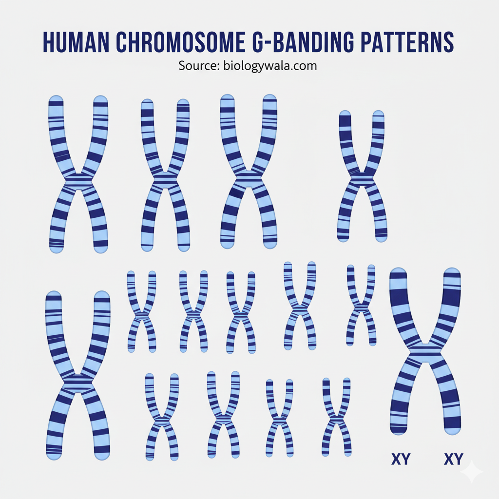
Chromosome banding techniques are staining methods that produce distinct light and dark bands along the length of chromosomes.
Each chromosome shows a unique banding pattern, allowing precise identification, structural analysis, and detection of abnormalities.
These patterns reflect differences in DNA composition, chromatin condensation, and base-pair richness (AT or GC content).
2. Principle
Banding depends on selective binding of dyes to regions of chromatin that differ in:
- Base composition (AT- or GC-rich regions)
- Degree of chromatin condensation
- Protein–DNA interactions
3. Major Types of Banding Techniques
| Type of Banding | Stain / Technique Used | Regions Highlighted | Key Features / Applications |
| G-banding (Giemsa banding) | Trypsin treatment followed by Giemsa stain | AT-rich regions | Produces alternating dark (heterochromatin) and light (euchromatin) bands; standard method for karyotyping. |
| Q-banding (Quinacrine banding) | Quinacrine fluorescent dye | AT-rich regions | Bands fluoresce under UV light; similar to G-banding but uses fluorescence microscopy. |
| R-banding (Reverse banding) | Heat treatment before Giemsa staining | GC-rich regions | Reverse of G-banding; highlights gene-rich, transcriptionally active areas. |
| C-banding (Constitutive heterochromatin banding) | Alkali and acid treatment before Giemsa staining | Centromeric and pericentromeric regions | Stains constitutive heterochromatin; useful for studying centromeres and repetitive DNA. |
| T-banding (Telomeric banding) | Modified R-banding | Telomeric ends | Stains telomeric regions intensely; reveals terminal chromosomal differentiation. |
| N-banding (Nucleolar organizer banding) | Silver nitrate (AgNO₃) stain | NOR (nucleolar organizer regions) | Identifies chromosomes involved in rRNA synthesis (satellite regions). |
4. Applications of Banding Techniques
- Karyotype identification – distinguishing individual chromosomes and homologous pairs.
- Detection of chromosomal abnormalities – such as deletions, duplications, inversions, and translocations.
- Comparative cytogenetics – studying chromosomal homology across species.
- Evolutionary studies – understanding chromosomal rearrangements during evolution.
- Medical diagnostics – identifying genetic disorders (e.g., Down, Turner, and Cri-du-chat syndromes).
Banding pattern analysis revolutionized cytogenetics by providing chromosomal fingerprints for each species.
It enables detailed structural and functional mapping of chromosomes, linking morphological features to genetic function and evolution.
https://biologywala.com/karyotype-analysis-and-evolution
Join SACHIN’S BIOLOGY on Instagram or Facebook to receive timely updates and important notes about exams directly on your mobile device. Connect with Mr. Sachin Chavan, the founder of Sachin’s Biology and author of biologywala.com, who holds an M.Sc., NET JRF (AIR 21), and GATE qualifications. With SACHIN’S BIOLOGY, you can have a direct conversation with a knowledgeable and experienced professional in the field of biology. Don’t miss out on this opportunity to enhance your exam preparation!


