Molecular Organisation of Centromere and Telomere

Chromosomes are the physical carriers of genetic material, and their functional integrity during the cell cycle depends critically on two specialized regions:
- the centromere, essential for chromosome movement and segregation, and
- the telomere, essential for chromosome stability and replication.
Although both appear as specific regions under the microscope, modern molecular biology has revealed that they are highly organized DNA–protein complexes with unique sequence motifs and epigenetic characteristics.
2. Centromere – Structure and Organisation
2.1 Definition
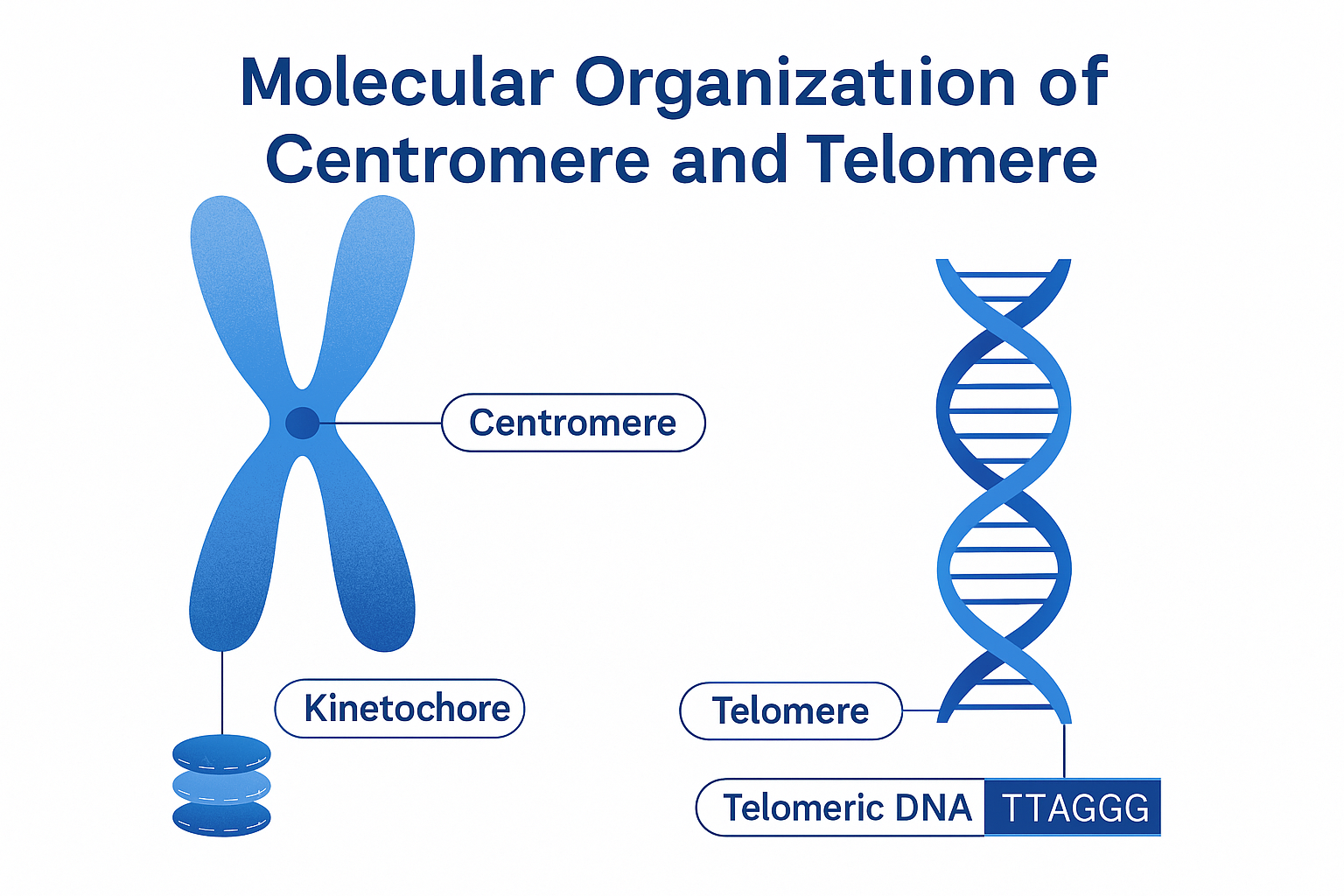
The centromere (primary constriction) is the chromosomal region where kinetochore proteins assemble, allowing attachment of spindle microtubules during mitosis and meiosis.
It ensures accurate distribution of chromosomes to daughter cells.
2.2 Position and Types
Based on the position of the centromere, chromosomes are classified as:
| Type | Position | Shape during Anaphase |
| Metacentric | Middle | V-shaped |
| Submetacentric | Slightly off-center | L-shaped |
| Acrocentric | Near one end | J-shaped |
| Telocentric | At the end | i-shaped |
| Holocentric | Diffuse (along entire length) | Parallel movement (e.g., Caenorhabditis elegans) |
2.3 Molecular Composition
The centromere is a complex DNA–protein domain containing:
A. DNA Components
- Repetitive DNA Sequences:
- Centromeric DNA consists mainly of satellite DNA – highly repetitive, non-coding sequences.
- In humans: α-satellite DNA (171 bp repeats) forms the major component.
- In plants: CentO (155 bp) repeats in rice, pAL1 in maize, and TrsA in Arabidopsis.
- CEN DNA (Centromeric Core DNA):
- A small region (120–150 bp in yeast) responsible for kinetochore binding.
- Yeast has a “point centromere”, while higher eukaryotes have “regional centromeres” several megabases long.
- Centromeric Heterochromatin:
- Rich in AT base pairs, tightly packed, late-replicating.
- Contains transposon-like elements and tandem repeats.
B. Protein Components
The centromeric chromatin differs from normal nucleosomes.
It contains specialized histone variants and kinetochore proteins:
| Protein | Function |
| CENP-A (CENH3 in plants) | A centromere-specific variant of histone H3 that replaces canonical H3; imparts unique chromatin identity. |
| CENP-B, CENP-C, CENP-E | Structural proteins forming the kinetochore complex. |
| CENP-T, CENP-N | Link centromeric DNA to microtubules. |
| Topoisomerase II | Relieves torsional stress during condensation. |
| Cohesins | Hold sister chromatids together until anaphase. |
2.4 Kinetochore Organisation
The kinetochore is a trilaminar proteinaceous structure formed on the centromere during cell division.
| Layer | Composition and Function |
| Inner plate | Attached directly to centromeric DNA containing CENP-A chromatin. |
| Middle layer | Contains structural proteins (CENP-C, CENP-H) forming the core scaffold. |
| Outer plate | Binds to microtubules through motor proteins like dynein and kinesin. |
Each kinetochore binds 20–40 microtubules in human cells, depending on the chromosome size.
2.5 Epigenetic Identity of the Centromere
Centromere identity is epigenetically inherited — not strictly determined by DNA sequence but by:
- Presence of CENP-A nucleosomes,
- Histone modifications (H3K9me3, H4K20me3), and
- DNA methylation patterns.
This ensures centromere function is maintained across generations, even if the sequence composition varies.
2.6 Function of Centromere
- Attachment site for spindle fibres via kinetochores.
- Ensures equal segregation of chromosomes during cell division.
- Maintains sister chromatid cohesion until anaphase.
- Involved in chromosome movement through motor proteins.
- Prevents chromosome breakage during segregation.
3. Telomere – Structure and Organisation
3.1 Definition
A telomere is a specialized repetitive DNA–protein complex present at the ends of linear chromosomes, providing stability, replication completeness, and protection against degradation.
Telomeres were first described by Müller (1938) and McClintock (1941), who observed that chromosome ends possess unique structures preventing fusion.
3.2 Molecular Composition of Telomeres
A. DNA Components
- Repetitive Sequences:
- Consist of short tandem repeats rich in guanine (G-rich strand).
- Common motif:
- Humans and vertebrates: (TTAGGG)ₙ
- Plants (Arabidopsis): (TTTAGGG)ₙ
- Tetrahymena: (TTGGGG)ₙ
- Single-Stranded Overhang:
- The G-rich 3′ overhang (~100–200 nucleotides) folds back and invades the double-stranded region, forming a T-loop (telomeric loop) and a D-loop (displacement loop) structure for protection.
B. Protein Components
Telomeric DNA is coated by specific protein complexes forming the shelterin complex (in vertebrates) or telosome (in plants and yeast).
| Protein | Role |
| TRF1, TRF2 (Telomeric Repeat-binding Factors) | Bind double-stranded telomeric DNA; maintain T-loop formation. |
| POT1 (Protection of Telomeres 1) | Binds to single-stranded G-rich overhang. |
| TIN2, TPP1, RAP1 | Link TRF1/2 and POT1, stabilizing the telomere complex. |
| Telomerase enzyme | Adds telomeric repeats to the 3′ ends during replication. |
3.3 Telomerase and Replication of Telomeres
Because DNA polymerase cannot fully replicate the 3′ ends of linear chromosomes (the “end-replication problem”), cells use telomerase, a ribonucleoprotein reverse transcriptase enzyme.
- Telomerase components:
- TERT (Telomerase Reverse Transcriptase) — protein component.
- TERC (Telomerase RNA Component) — RNA template (e.g., 3′-CAAUCCCAAUC-5′ in humans).
Function:
Telomerase extends the G-rich strand by adding repeats complementary to its RNA template, preventing telomere shortening during successive cell divisions.
3.4 Functions of Telomeres
- Chromosome End Protection:
- Prevents chromosome ends from being recognized as DNA breaks.
- Prevention of End-to-End Fusions:
- Shelterin complex inhibits DNA repair machinery from ligating ends.
- Complete Replication:
- Telomerase ensures full replication of chromosome ends.
- Nuclear Architecture:
- Anchors chromosomes to the nuclear periphery.
- Aging and Disease:
- Telomere shortening is associated with cellular aging (senescence), cancer, and genomic instability.
3.5 Comparison: Centromere vs. Telomere
| Feature | Centromere | Telomere |
| Location | Primary constriction (middle region) | Terminal ends of chromosome |
| DNA Type | Satellite repetitive DNA | Tandem G-rich repeats |
| Proteins | CENP complex (CENP-A, CENP-B, etc.) | Shelterin complex (TRF1, TRF2, POT1, etc.) |
| Function | Kinetochore formation, chromosome segregation | End protection, replication completion |
| Replication Timing | Late S-phase | Late S-phase (with telomerase) |
| Chromatin Type | Constitutive heterochromatin | Specialized heterochromatin |
| Epigenetic Marker | CENP-A histone variant | G-quadruplex DNA, telomerase activity |
3.6 Structural Overview Diagram (Conceptual)
Chromosome:
[Telomere] — [Euchromatin] — [Centromere] — [Euchromatin] — [Telomere]
Telomere: (TTAGGG)n repeats → T-loop → Shelterin complex
Centromere: α-satellite repeats → CENP-A nucleosomes → Kinetochore attachment
4. Summary
| Aspect | Centromere | Telomere |
| Primary role | Chromosome segregation | Chromosome end protection |
| DNA nature | AT-rich satellite repeats | GC-rich tandem repeats |
| Protein complexes | Kinetochore, CENPs | Shelterin, telomerase |
| Histone modification | H3 variant CENP-A | Histone H3K9me3, TERRA RNA |
| Dynamic function | Microtubule attachment | Replication and aging regulation |
Both centromeres and telomeres are specialized chromosomal domains essential for genome integrity.
- The centromere ensures faithful segregation of chromosomes through kinetochore formation and spindle attachment.
- The telomere maintains chromosome stability, prevents end-to-end fusion, and regulates cellular lifespan through controlled telomerase activity.
Together, they represent the molecular guardians of chromosome architecture — safeguarding genetic continuity across generations.
https://biologywala.com/download-complete-notes-on-chromatin-organisation-pdf
Join SACHIN’S BIOLOGY on Instagram or Facebook to receive timely updates and important notes about exams directly on your mobile device. Connect with Mr. Sachin Chavan, the founder of Sachin’s Biology and author of biologywala.com, who holds an M.Sc., NET JRF (AIR 21), and GATE qualifications. With SACHIN’S BIOLOGY, you can have a direct conversation with a knowledgeable and experienced professional in the field of biology. Don’t miss out on this opportunity to enhance your exam preparation!


