CHROMOSOME: Types, Parts, structure Disorders

CHROMOSOME:
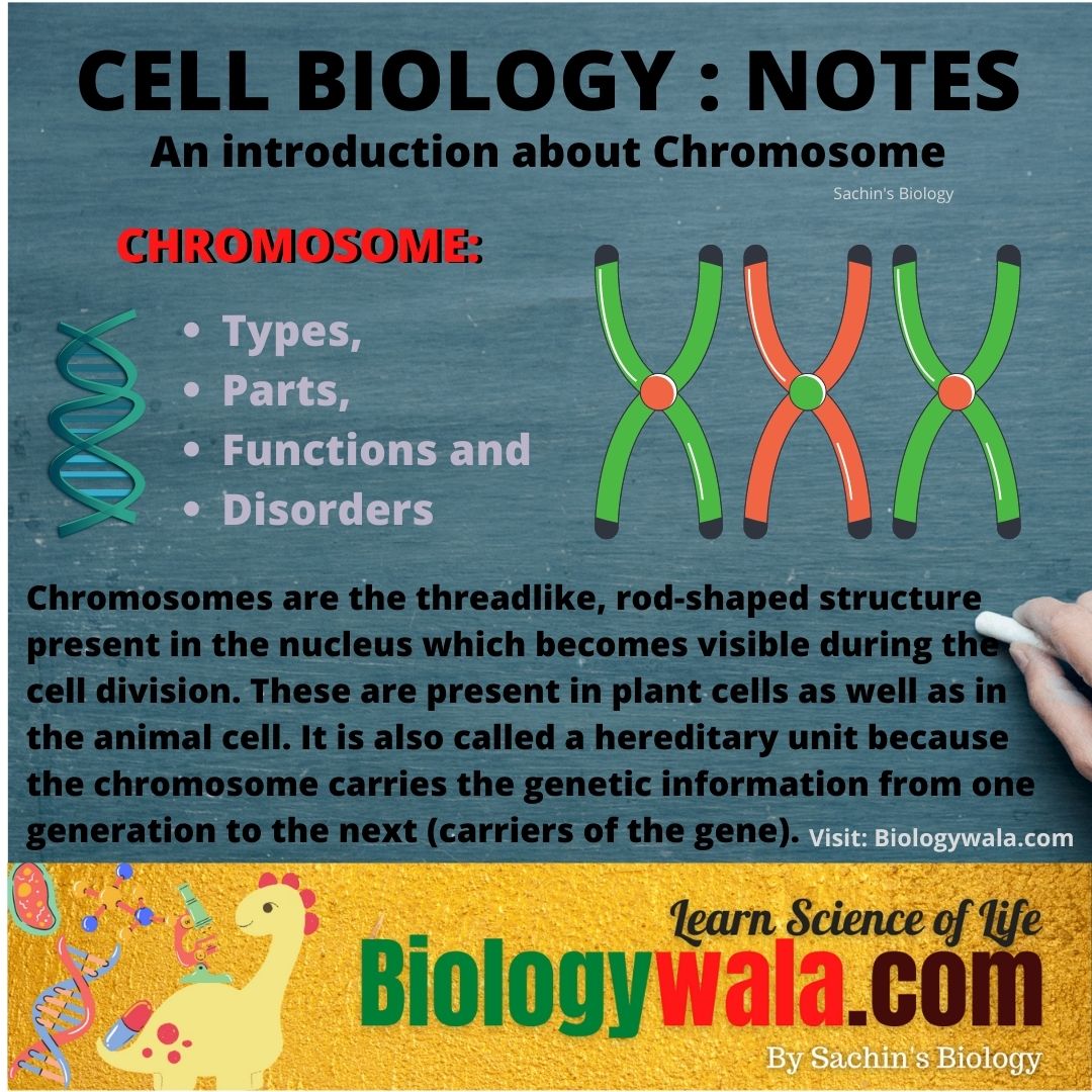
Chromosomes are the threadlike, rod-shaped structure present in the nucleus which becomes visible during the cell division. These are present in plant cells as well as in the animal cell. It is also called a hereditary unit because the chromosome carries the genetic information from one generation to the next (carriers of the gene).
The chromosome was first discovered by Strasburger in 1875, the term chromosome was first used by Waldeyer in 1888 (chromo=colour; soma=body) due to their marked affinity for the basic dyes.
In humans, each cell normally contains 23 pairs of chromosomes, for a total of 46. Out of 23 pairs, the twenty-two pairs are called autosomes, these look the same in both males and females. The 23rd pair is the sex chromosomes, which differ between males and females. In the female X-chromosome is present and in the male Y-chromosome is present.
Parts of chromosomes and their functions:
The chromosome has 6 main parts;
- Pellicle and matrix 2. Chromatids, chromonema and chromomeres 3. Centromeres 4. Secondary construction 5. Satellite 6. Telomere.
Part # 1. Pellicle and matrix:
A membrane that surrounds each chromosome is called a pellicle and the jelly-like substance present inside the membrane is called a matrix. This matrix is present in the chromonemata. This matrix provides the basic shape and structure of the nucleus.
Part # 2. Chromatids, chromonema, and chromomeres:
Chromatid is the structural and functional unit of the chromosomes. The chromosome is made up of chromatin, and chromatin is the most important constituent of the cell nucleus. The structure of the chromosome is best studied in late prophase, metaphase and anaphase. During the early prophase, a chromosome appears to consist of spirally coiled thin or filamentous long continuous structures are called chromonema.
The chromosomes of some of the species show the small bead-like structure is called chromomeres. The structure of the chromomeres in the chromosome is constantly based on which a chromosome can be differentiated from one another chromosome. These chromosomes are responsible to carry genes during the inheritance.
Part # 3. Centromere or primary constriction or kinetochore
The two chromatids are joined together at a point along their length which is said as centromere or primary constriction or kinetochore. It is the clear and non-stainable part of the chromosome.
It is the construction towards the upper end of the chromosome. The position of the centromere on a chromosome may vary in different chromosomes.
The parts of the chromosome on each side of the centromere are called an arm.
The function of the centromere is its plays important role in directing the process of movement during the cell division. It performs two roles i.e., holding the two chromatids and binding the spindle fibres. It may be single at one stage double or four-stranded at another stage like meiosis, prophase 1 pachytene.
Depending upon the position of centromere and comparative length of two arms of the chromosome, chromosome may be of following types;
1: Acrocentric
Acrocentric chromosomes have the centromere are almost at the one end
2: Metacentric chromosome
The metacentric chromosome has a centromere in the middle of the chromosome. It appears V-shaped structure and the arms are equal in length.
3: Sub-metacentric
Sub-metacentric chromosomes have a centromere slightly off-centre. The structure is L or J-shaped. The arms are unequal.
4: Telocentric
The ends of each chromosome are called telomeres. Functionally telomere is a complete end of chromosomes. It has the unique function of preventing chromosomes from attaching. The structure is I-shaped, single-arm.
5: Dicentric or polycentric :
The chromosome has two or more centromeres. It is formed through the fusion of two chromosome segments, each with a centromere resulting in the loss of acentric fragments and the formation of dicentric fragments.
6: Acentric:
When the chromosome lacks the centromere, it is called an acentric chromosome.
Part # 4 . Secondary constructions
In addition to the centromere, many chromosomes have one or more secondary constructions. It is also said as the nucleolar organising region because they are necessary for the formation of the nucleolus. The function of this secondary construction is, it marks the site of a nucleolar organizer region (NOR), a region containing multiple copies of the 18s and 28s ribosomal genes that synthesize ribosomal RNA required by ribosomes. The nuclear organizer represents about 0.3% of the total amount of nuclear DNA.
Part # 5. Satellite
The secondary construction usually appears as a single small body or a pair of such small bodies attached to the remainder of the chromosome by a slender thread. These chromosome bodies are known as satellites. Satellites are elongated, round in shape and variable in size. The chromosome with a satellite is referred to as SAT-chromosomes. There are at least two SAT-chromosome in each diploid nucleus and many polyploid species also have only two SAT-chromosomes. For example in wheat hexapods.
Part # 6. Telomere
The end of each chromosome is called telomeres. Functionally telomere is completed end of the chromosome. The function of chromosomes is unique, it prevents chromosomes from attaching. A broken chromosome will not be attached to a telomere unless that telomere is also fractured.
Telomere is the specially modified portion of the chromosomes for attachment to the nuclear membrane.
Types of chromosomes:
Chromosomes are of two types Autosomes and Sex chromosome {allosomes}
Autosomes:
Autosomes control characters other than the sex characters or carry the gene for somatic characters.
Sex chromosome:
Chromosomes are involved in sex determination. For example- humans have two sex chromosomes X and Y. Females have two X chromosomes in diploid cells and males have X and Y chromosomes.
Disorders caused by chromosomal abnormalitiesChromosome Disorder in humans | Chromosomal disorders list:
- Cri du chat syndrome : 5p minus syndrome (partial deletion of short arm of syndrome.
- XYY syndrome and XXX syndrome .
- Downs syndrome : trisomy 21
- Patau syndrome: trisomyn 13
- Edwards syndrome : trisomy 18
- Wolf-Hirschhorn syndrome : deletion disorder
- Klinefelters syndrome: presence of additional X chromosome in males.
- Turner syndrome: presence of only a single X chromosome in females.
Disorders of Sex Development.
Down Syndrome.
Klinefelter Syndrome.
Genetic Disorder.
Congenital Malformation.
Aneuploidy.
Summary
Chromosomes are generally decondensed during interphase so that the details of their structure are difficult to visualize. Notable exceptions are the specialized lampbrush chromosomes of vertebrate oocytes and the polytene chromosomes in the giant secretory cells of insects. Studies of these two types of interphase chromosomes suggest that each long DNA molecule in a chromosome is divided into a large number of discrete domains organized as loops of chromatin that are compacted by further folding. When genes contained in a loop are expressed, the loop unfolds and allows the cell’s machinery access to the DNA.
Interphase chromosomes occupy discrete territories in the cell nucleus; that is, they are not extensively intertwined. Euchromatin makes up most of the interphase chromosomes and, when not being transcribed, it probably exists as tightly folded fibres of compacted nucleosomes. However, euchromatin is interrupted by stretches of heterochromatin, in which the nucleosomes are subjected to additional packing that usually renders the DNA resistant to gene expression.
Heterochromatin exists in several forms, some of which are found in large blocks in and around centromeres and near telomeres. But heterochromatin is also present at many other positions on chromosomes, where it can serve to help regulate developmentally important genes. The interior of the nucleus is highly dynamic, with heterochromatin often positioned near the nuclear envelope and loops of chromatin moving away from their chromosome territory when genes are very highly expressed.
This reflects the existence of nuclear subcompartments, where different sets of biochemical reactions are facilitated by an increased concentration of selected proteins and RNAs. The components involved in forming a subcompartment can self-assemble into discrete organelles such as nucleoli or Cajal bodies; they can also be tethered to fixed structures such as the nuclear envelope.
During mitosis, gene expression shuts down and all chromosomes adopt a highly condensed conformation in a process that begins early in the M phase to package the two DNA molecules of each replicated chromosome as two separately folded chromatids.
The condensation is accompanied by histone modifications that facilitate chromatin packing, but satisfactory completion of this orderly process, which reduces the end-to-end distance of each DNA molecule from its interphase length by an additional factor of ten, requires additional proteins.
What are the parts of chromosomes?
There are total main 6 types of the chromosome
Pellicle and matrix 2. Chromatids, chromonema and chromomeres 3. Centromeres 4. Secondary construction 5. Satellite 6. Telomere.
What are chromosomes mainly made of?
A chromosome is a thread-like structure it is made up of protein and a single molecule of deoxyribonucleic acid (DNA).
This unique structure of chromosome holds DNA tightly bound around spool-like proteins, called histones.
How many genes are in a chromosome?
The chromosome contains hundreds to thousands of genes. This gene carries the instructions for making proteins. Each of the estimated 30,000 genes in the human genome makes an average of three proteins.
How many chromosomes does a human have?
46 Chromosomes in 23 pairs.
I hope you like this article. If you think there should be some additional information in the article that need to be added then let us know in the comment box. You can also suggest topic for next article.
You will also like :
[FREE [Download] Campbell Biology Concepts & Connections 9th Edition pdf book
[Downlaod] Free Campbell Essential Biology 7th Edition PDF Book
If you want important notes and updates about exams on your mobile then you can join SACHIN’SBIOLOGY on Instagram or Facebook and can directly talk to the founder of Sachin’s Biology and Author of biologywala.com Mr.Sachin Chavan M.Sc. NET JRF (AIR 21) GATE!
![[PDF] 10 Rod Cells and cone cells Differences | Rod Cells Vs cone cells Biology PDF Notes](https://biologywala.com/wp-content/themes/hueman/assets/front/img/thumb-medium-empty.png)
